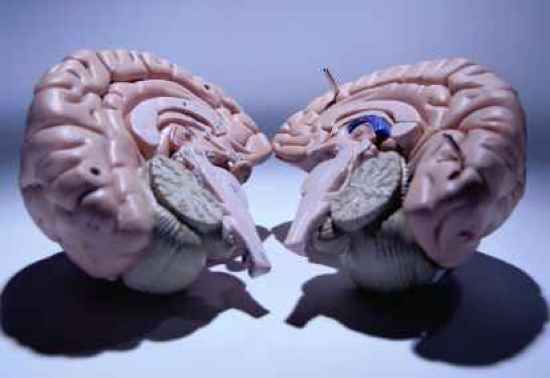
Location of Damage: Corpus Callosum Peter began to suffer from complex partial seizures at the age of 8. His seizures were severe, despite taking antiepileptic medication; so, at the age of 20, after struggling with his condition for over 12 years, he and his doctors agreed to have him undergo a commissurotomy. The commissurotomy involved a surgical incision of Peter’s corpus callosum. The corpus callosum is the major cerebral nerve tract that connects the brain’s right and left cerebral hemispheres together; and therefore, serves as the major communication pathway for intrahemispheric signals. Removal or damage to one’s corpus callosum would result in impaired communication between the two sides of the brain. On one hand, Peter’s surgery was a success, as it did end up attenuating the magnitude of his seizures. On the other hand, however, Peter was left much different than before. For example, he was not able to respond with the left side of his body to verbal input. If asked to “Stand like a Boxer,” his left side would sag and appear lackluster, while his right side would behave appropriately. Because his brain had, literally, been split into two separate pieces, his left and right sides were often behaving like two separate people. He complained that his left hand would turn off television shows that he was enjoying, that his left leg would not always walk in the intended direction, and that his left arm would occasionally begin to fight with the right side of his body. It is clear from Peter’s case that the brain works best when it is capable of functioning as a single, cohesive unit, as opposed to multiple ones.
Location of Damage: Primary Visual Cortex (Occipital Lobe) In everyday thinking, perceiving and being consciously aware of something are one and the same; they are inseparable processes. We readily assume that someone who has seen something will always be able to acknowledge, and verbally describe what that object was. This ability to see and perceive consciously is something we, in fact, take for granted. The case of D.B. is a prime example of this. D.B. suffers from a neurological impairment known as blindsight, which is the inability for one to consciously perceive something that one can see. D.B. has very good eyesight; in fact, his sight is better than the average normal person. What makes him different is that he suffers from damage to his primary visual cortex, a part of the brain’s occipital lobe. Despite the fact that his brain damage has caused him to lose awareness of “seeing” in his left visual field, evidence has been obtained for the following: (a) he is capable of reaching for visual stimuli in his left field with considerable accuracy; (b) he can differentiate the orientation of a vertical line from a diagonal or horizontal line; (c) he can tell the difference between the letters “X” and “O” when they are shown in his field of blindness. It goes without saying that his doctors questioned him repeatedly about the vision in his left half-field, only to respond that he saw nothing at all. When he was shown his results (through his intact, right visual field), he expressed great surprise and insisted that he was just “guessing.” D.B. was astonished when shown video footage of himself accurately reaching and judging the orientation of lines. Even though he could not consciously register what had been presented in his ‘blind’ visual field, he still possessed the ability to ‘see’ in a sense, hence the term ‘blindsight’.
Location of Damage: Fusiform Face Area We constantly rely on our ability to discern and recognize people by the nature of their faces. Imagine what life would be like if everyone you knew wore a paper bag over their heads, so as to obfuscate their identities. Weird, isn’t it? This is exactly the life R.P. experiences everyday. R.P. suffers from prosopagnosia, which is an inability to recognize faces. In fact, prosopagnosia is only one of many visual agnosias (an agnosia is a failure of recognition). Other visual agnosias include: movement agnosia, object agnosia and color agnosia. R.P. can usually recognize a face is a face, but generally cannot tell exactly whose face it is. R.P. does not report any other recognition problems, and like D.B. in the previous entry, has superb vision. Prosopagnosics often report seeing a jumble of individual facial parts that are not bound, or fused, into an easily recognizable whole. In some extreme cases, they cannot even recognize themselves. Prosopagnosia is associated with damage to the boundary between the brain’s occipital and temporal lobes, a region better known as the fusiform face area.
Location of Damage: Complete Removal of Left Hemisphere Before contracting her illness, N.I. was a perfectly normal girl. Unfortunately, at the age of 13, she began to experience convulsions, along with deteriorated speech and motor abilities. CT scans revealed ischemic (lack of blood flow) brain damage to her left hemisphere. N.I. was still experiencing symptoms two years after the onset of her illness, and her right limbs had become paralyzed. In an attempt to relieve these symptoms, a total left hemispherectomy (removal of a hemisphere in the brain) was performed. Following this procedure, her seizures subsided completely. Sadly, however, her surgery had taken away her ability to read correctly. (Most people, including N.I., use their left hemispheres for language related tasks) Post surgery, N.I. was able to recognize letters, but was totally incapable of translating them into sounds. She could read concrete familiar words (ie: fan), but could not pronounce even simple nonsense words (ie: neg). Her reading errors indicate that she reads on the basis of meaning and appearance of words rather than by translating the individual letters into sounds. For instance, when shown the word “fruit” she would respond by saying: “juice.” Her responses are similar to people with a disorder known as ‘deep dyslexia’, which is an inability to apply rules of pronunciation while reading. Deep dyslexics can still pronounce familiar words based on their specific memories of them, but similar to N.I., they cannot pronounce nonsensical words, or words they are unfamiliar with.
Location of Damage: Right Occipital and Parietal Lobes W.M. suffered a stroke in his right posterior cerebral artery. The stroke caused extensive damage to his right occipital and temporal lobes, which resulted in severe left hemianopsia. Hemianopsia is a condition where a scotoma (a blind spot in your field of vision) covers half of your visual field. In essence, W.M. was virtually blind in his left visual field. When tested with his left hand on his lap, W.M. detected 97.8% of the stimuli presented to his right visual field, but only 13.6% of those presented on the left. What makes his case interesting is that when he was tested with his left hand extended into his left visual field, his ability to detect stimuli improved markedly. For some reason, his brain allowed him to see those objects that were near his left hand, even if his hand was within the region of his scotoma (his left visual field). Remarkably, this area of improved performance was expanded even further when he held a tennis racket in his extended left hand. W.M.’s case is a testament to the durability of the human brain. Even when damaged, the brain often develops abilities to adapt to, and work around, its impairment.
Location of Damage: Unknown (most likely caused by a genetic defect; one that disrupts the synthesis of sodium ion channels within the brain’s anterior cingulate cortex) The perception of pain is highly paradoxical in nature. It’s tough to live ‘with’ and it’s tough to live ‘without’. It’s funny how an experience that seems in every respect to be so bad is, in fact, extremely important for our survival. Pain is needed because it warns us to stop engaging in potentially harmful activities. The importance of pain is best illustrated by cases in which people experience no pain; the case of Miss C. is such an example. Miss C., a highly intelligent young lady who was normal in every respect, save for the fact that she had never felt pain, not even in the slightest form. When she was a child, she had bitten off the tip of her tongue while chewing food. The marks of third degree burns were scattered across her body after she had burned herself while unknowingly kneeling on a radiator. She could not feel the shock of electricity flowing through her body, and felt no pain to both extremely hot, or cold water. Furthermore, she did not experience a change in physiological responses when subjected to such extremes (eg. no change in blood pressure, respiration, heart rate, etc.). When asked, Miss C. could not remember the last time she had coughed or sneezed. Moreover, a gag reflex could only be brought forth with great difficulty and her corneal reflexes (reflexive blinking) were entirely absent. She failed to shift her weight when standing upright, and did not turn over in her sleep (these actions are necessary to prevent inflammation of the joints). Sadly, Miss C. passed away at the age of 29 due to complications from infections, skin and bone trauma.
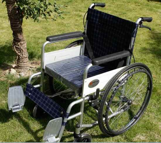
Location of Damage: Right Posterior Parietal Cortex After her stroke, Mrs. S. had been unable to recognize or respond to things on her left (this would include both external objects, and parts of her own body). For example, she often put makeup on the right side of her face but not her left. Mrs. S was suffering from contralateral neglect, which is a disturbance of one’s ability to respond to stimuli on the side of the body opposite (contralateral) to the site of brain damage. In the case of Mrs. S, her stroke had damaged her brain’s right posterior parietal lobe and, thus, she was plagued by left-side contralateral neglect. This disability, as one might imagine, created many problems for her. One particularly troublesome problem was that she had difficulty getting enough to eat. When a plate full of food was placed in front of her, she could only see the food on the right side of the plate and, therefore, only ate that much, despite being very hungry. Over time, Mrs. S. developed a clever way of getting more food if she was still hungry. While in a wheelchair, she would turn it around to the right in a full circle and stop when the remaining half of her meal would become visible. She would eat that, then turn once more until the remaining half of the previous portion came into view, and again, eat that. She would repeat this process until she was content or the plate became emptied, whichever came first.
Location of Damage: Medial Diencephalon (Medial Thalamus and Medial Hypothalamus) The final three entries of this list will deal with, arguably, the most notorious of psychopathological ailments, namely, amnesia (an impairment of memory). Let us begin by examining the unfortunate case of Jimmie G. Jimmie G. was a gregarious 49-year-old man, who enjoyed talking about his experiences as a schoolboy and about his time in the navy. He was intelligent, and possessed superior math and science abilities. Given this description, it would be puzzling for anyone to believe that he was a resident of a neurological ward. A glimpse of his problem could be caught when one paid careful attention to his description about his past. When recounting his experiences as a schoolboy, he used past tense; however, when recalling his experiences from the navy, he switched to present tense. Even stranger was the fact that he never talked about anything that happened to him after his time in the navy. Jimmie G. believed that he was a 19-year-old young man still serving his country. When shown his reflection, he would become confused and disgruntled. If you were to leave his presence only to come back a few minutes later, Jimmie G. would greet you as if he had never seen you before. When asked where he thought he was, Jimmie would respond by saying that it looked like he was in a hospital, and he was afraid that he had been admitted because he was sick, but didn’t know it. Additional testing revealed that he was suffering from Korsakoff’s syndrome, which is a disorder displayed in people who have consumed large amounts of alcohol. (Jimmie G. was indeed an alcoholic as a young man). His amnesia is attributable to the brain damage caused by thiamine deficiency (vitamin B1), most often seen displayed in chronic alcoholics. In its advanced stages, like that of Jimmie G’s case, Korsakoff’s syndrome produces profound amnesic effects.
Location of Damage: Diffuse damage; mainly the medial temporal lobes K.C. had been involved in a motorcycle accident, in 1981. As a result, he suffered diffuse brain damage and severe amnesia. Despite his memory impairment, K.C.’s other cognitive abilities remained remarkably normal. He has no difficulty concentrating; he plays chess, the organ, various card games and his reasoning abilities are good. His knowledge of mathematics, science, history, geography and other subjects learned in school are also good. He has good retention for the many facts he had learned in his early life, such as his birth date, his address when he was a child, the names of the schools he attended, etc. If you were paying close attention, you have probably noticed that all of his intact memories possess a single attribute. That is to say, all of his memories are factual in nature (these types of memories are called semantic memories). While his semantic knowledge remains intact, K.C. has severe amnesia for personal experiences. He cannot recall a single personal event for more than a minute. This inability to recall any events (episodes) which he experienced covers the span of his entire life. Despite these serious memory problems, K.C. has no difficulty holding a conversation, and his memory problems are much less obvious than one would expect. He does a very good job of using his ‘semantic memory’ during conversations. For example, when asked what his brother’s funeral was like, he would respond by saying “Sad, very sad.” However, K.C. could not recall ever even having a brother, let alone whether or not he was still alive. From his semantic memory, K.C. was able to retrieve the fact that funerals were sad events, and therefore, used this knowledge to answer the question accordingly. K.C. understands the concept of time, but cannot “time travel,” into either the past or the future. He cannot recall his past any better than he can envision his future. In effect, he cannot imagine what he will be doing for the rest of the day, the week, or his life.
Location of Damage: Removal of Both Medial Temporal Lobes (Loss of hippocampus, amygdala, and perirhinal cortex) The case of H.M. (Henry Molaison) is probably the most well known case on this list. Amidst all of its sadness, it has helped many scientists to advance the study of the nature of human memory. For those of you who are not familiar with it, here is his story: H.M. had suffered from severe epilepsy during the eleven years preceding his surgery. He experienced, on average, one generalized seizure each week, and many partial seizures each day, despite being on anticonvulsants. Results from an electroencephalography (EEG) test showed that his convulsions stemmed from the medial (inner) portions of his left and right temporal lobes. A decision was made to remove both of these lobes, in hopes of stopping the convulsions (the procedure performed on H.M. was called a bilateral medial temporal lobectomy). The surgery did help alleviate his epilepsy, eliminating the general seizures entirely, and curtailing the frequency of partial seizures to one or two per day. Be that as it may, H.M. was the last patient to receive a bilateral medial temporal lobectomy; this was because the procedure had been responsible for producing profound amnesic effects. After his surgery, H.M. developed severe anterograde amnesia. Anterograde amnesia is a form of amnesia that impairs one’s memory for any event after the onset of brain damage (in contrast to retrograde amnesia, which impairs one’s memory for any event before the occurrence of brain damage). It turns out that H.M.’s general intelligence was not affected by the surgery (he scored above average on standardized intelligence tests). Much like his intellect, his memory for events predating the surgery was largely intact. In addition, H.M.’s short-term anterograde memory had also remained normal (ie: if given a list of 6 digits and asked to repeat them, he would do so without difficulty). Sadly, however, H.M. had an almost total inability to form new long-term memories. Once he stopped thinking about an experience, it would be lost forever. H.M. could not recognize those who he had met after his surgery, and would require a reintroduction as though he had never seen them before. He spent most of his day doing crossword puzzles in the nursing home he was a resident of. His progress on a crossword puzzle was never lost because it was written down. H.M. was a prisoner in his own mind, suspended in time, for the rest of his life.
